Home
/ Anatomy Arteries In Neck, Human Anatomy Structure Of The Human Body Arteries Anatomy Medical Anatomy Anatomy Organs, In the neck, the carotid sheath (fibrous connective tissue) covers the common carotid artery, vagus nerve, and internal jugular vein.
Anatomy Arteries In Neck, Human Anatomy Structure Of The Human Body Arteries Anatomy Medical Anatomy Anatomy Organs, In the neck, the carotid sheath (fibrous connective tissue) covers the common carotid artery, vagus nerve, and internal jugular vein.
Anatomy Arteries In Neck, Human Anatomy Structure Of The Human Body Arteries Anatomy Medical Anatomy Anatomy Organs, In the neck, the carotid sheath (fibrous connective tissue) covers the common carotid artery, vagus nerve, and internal jugular vein.. The carotid arteries are the primary vessels supplying blood to the brain and face. While both right and left arteries run the same course in the neck, they have differing origins. There are several head and neck arteries: The subclavian arteries arise asymmetrically but follow similar courses. Is the primary blood supply to the face and superficial head;
Internal carotid artery ( segments) caroticotympanic artery. Arteria subclavia) is a major blood vessel located in the thorax that provides blood supply to the upper limb, while some of its branches participate in supplying the head and neck. Contains glands ( thyroid, parathyroid, and thymus ), the larynx, pharynx and trachea. The right and left subclavian arteries give rise to the thyrocervical trunk. Contains cervical vertebrae and postural muscles.
Carotid Arteries from www.thoughtco.com In basic terms, the neck (cervical spine) joins the shoulders and chest to the head. In the neck, the carotid sheath (fibrous connective tissue) covers the common carotid artery, vagus nerve, and internal jugular vein. The right and left subclavian arteries give rise to the thyrocervical trunk. All major arteries of the neck originate from the aortic arch via three main vessels: The vertebral arteries terminate by anastomosing together as the basilar artery. In the neck, each carotid artery branches into two divisions: Arteria subclavia) is a major blood vessel located in the thorax that provides blood supply to the upper limb, while some of its branches participate in supplying the head and neck. The right common carotid artery (rcca) originates in the neck from the brachiocephalic artery while the left common carotid artery (lcca) arises in the thorax from the arch of the aorta.
Cadaveric angiographic and dissection studies have demonstrated that the external and internal carotids are the main arterial sources for the head and…
The right and left subclavian arteries give rise to the thyrocervical trunk. In the neck, each carotid artery branches into two divisions: In the neck, carotid arteries are contained within the carotid sheath posterior to the sternocleidomastoid muscle (one of the largest muscles). This section of the website will explain large and minute details of arterial anatomy of neck. Internal carotid artery ( segments) caroticotympanic artery. The content of the neck is grouped into 4 neck spaces, called the compartments. The right common carotid artery (rcca) originates in the neck from the brachiocephalic artery while the left common carotid artery (lcca) arises in the thorax from the arch of the aorta. Contains cervical vertebrae and postural muscles. The left common carotid comes directly off the aortic arch, while the right common carotid comes from the brachiocephalic. At the root of the neck the right internal jugular vein is placed at a little distance from the common carotid artery, and crosses the first part of the subclavian artery, while the left internal jugular vein usually overlaps the common carotid artery. The neck is one of the most complex and intricate structures in our body and includes the spinal cord, which sends messages from the brain to the rest of the body. Arteria subclavia) is a major blood vessel located in the thorax that provides blood supply to the upper limb, while some of its branches participate in supplying the head and neck. The head and neck receive the majority of blood through the carotid and vertebral arteries.
Through their course, they give off several meningeal, muscular and spinal branches for the nearby structures. Arteria subclavia) is a major blood vessel located in the thorax that provides blood supply to the upper limb, while some of its branches participate in supplying the head and neck. Is the primary blood supply to the face and superficial head; Contain the common carotid artery, internal. The vertebral arteries ascend through the neck inside the transverse foramina of the cervical vertebrae, all the way to the brain.
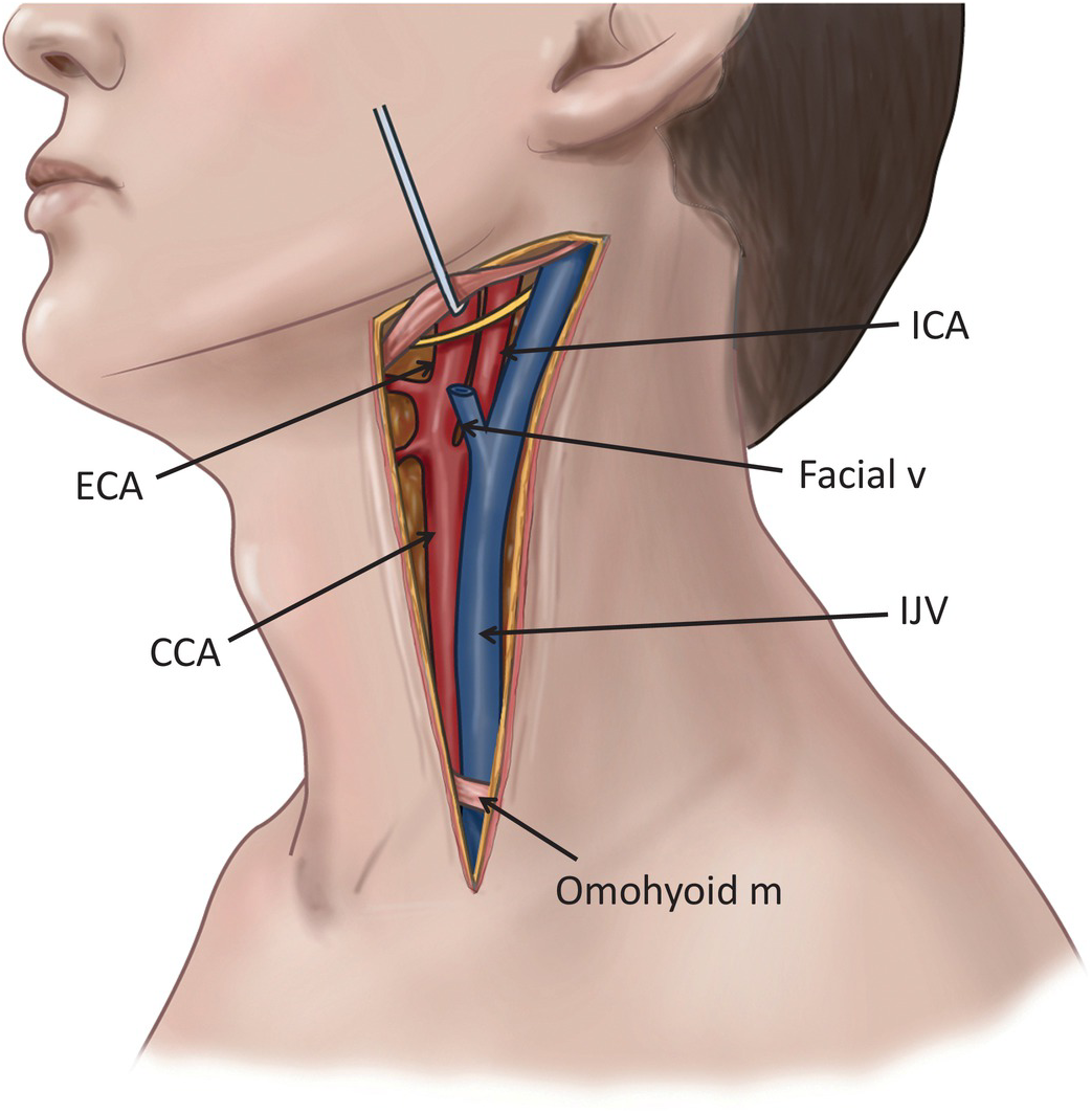
Carotid Artery And Internal Jugular Vein Injuries Chapter 8 Atlas Of Surgical Techniques In Trauma from static.cambridge.org There are several head and neck arteries: The neck is the area between the skull base and the clavicles. A large artery that arises on each side of the neck, the common carotid artery is the primary source of oxygenated blood for the head and neck. Cadaveric angiographic and dissection studies have demonstrated that the external and internal carotids are the main arterial sources for the head and… Through their course, they give off several meningeal, muscular and spinal branches for the nearby structures. In the neck, each carotid artery branches into two divisions: The carotid and vertebral arteries which travel through the area carry high. The left common carotid comes directly off the aortic arch, while the right common carotid comes from the brachiocephalic.
The brachiocephalic trunk, left common carotid (cca), and left subclavian arteries.
The first branch of the thyrocervical trunk is the inferior thyroid artery. In the neck, the carotid sheath (fibrous connective tissue) covers the common carotid artery, vagus nerve, and internal jugular vein. Contains glands ( thyroid, parathyroid, and thymus ), the larynx, pharynx and trachea. The anatomy of neck arteries, normal variations, and anastomoses between different arteries is discussed in this chapter. There are two carotid arteries, one on the right and one on the left. Contains cervical vertebrae and postural muscles. The neck is the area between the skull base and the clavicles. From this trunk, several vessels arise, which go on to supply the neck. The neck is one of the most complex and intricate structures in our body and includes the spinal cord, which sends messages from the brain to the rest of the body. The right common carotid artery (rcca) originates in the neck from the brachiocephalic artery while the left common carotid artery (lcca) arises in the thorax from the arch of the aorta. Common carotid artery the right and left common carotid arteries (cca) course superiorly on both sides of the neck lying within the respective carotid space, anteromedial to the internal jugular veins and accompanied by the vagus nerve. In basic terms, the neck (cervical spine) joins the shoulders and chest to the head. In the neck and head exterior to the skull, the external carotid artery provides blood flow to the skin, muscles, and organs.
The right common carotid artery (rcca) originates in the neck from the brachiocephalic artery while the left common carotid artery (lcca) arises in the thorax from the arch of the aorta. The vertebral arteries ascend through the neck inside the transverse foramina of the cervical vertebrae, all the way to the brain. The brachiocephalic trunk, left common carotid (cca), and left subclavian arteries. All major arteries of the neck originate from the aortic arch via three main vessels: Ophthalmic a., posterior communicating a., anterior cerebral a., middle cerebral a.
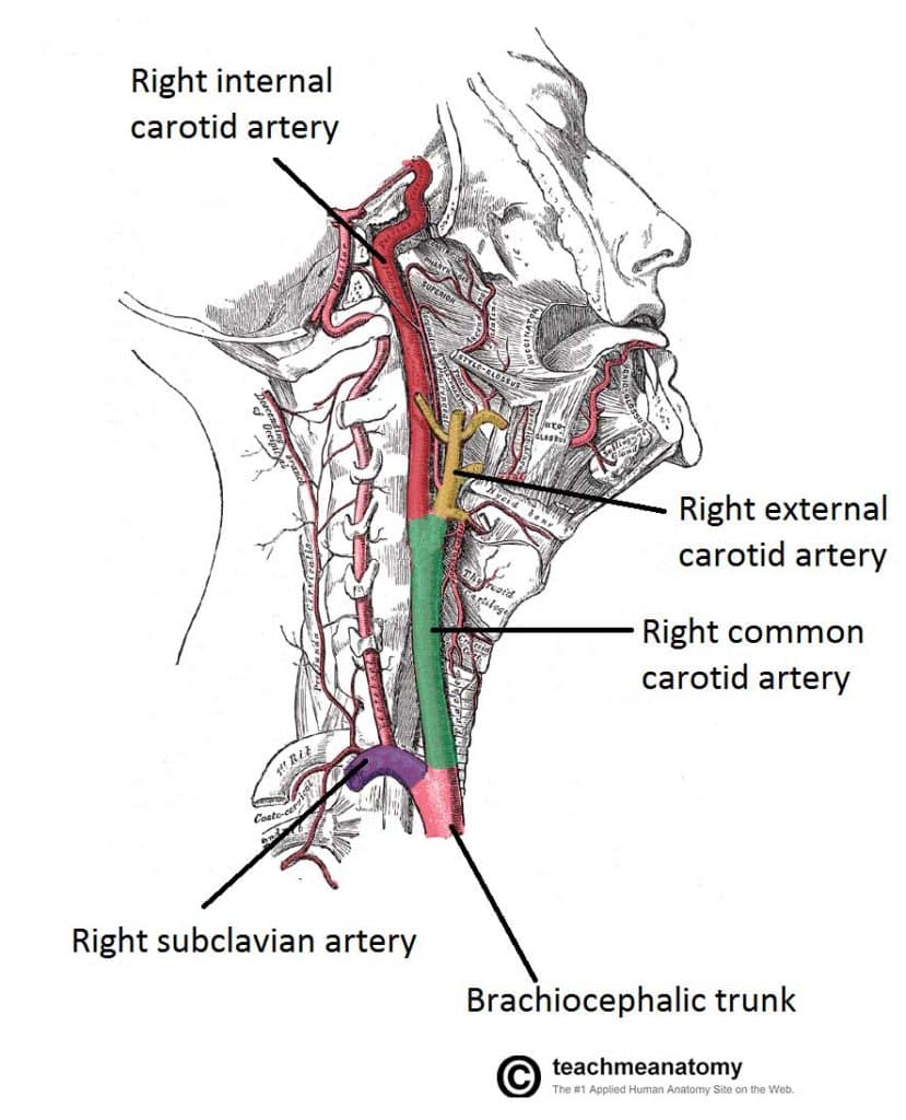
Major Arteries Of The Head And Neck Carotid Teachmeanatomy from teachmeanatomy.info The right common carotid artery is a branch of the brachiocephalic artery while the left common carotid arises from the arch of the aorta. The subclavian arteries arise asymmetrically but follow similar courses. The neck is supplied by arteries other than the carotids. The first branch of the thyrocervical trunk is the inferior thyroid artery. Contains cervical vertebrae and postural muscles. The vertebral arteries is a main division of the subclavian artery. The arteries of the head and neck are branches of the common carotid and subclavian arteries. Right common carotid artery arises from the brachiocephalic artery.
Left and right common carotid arteries supply the head and neck with oxygenated blood.
The vein is the most lateral structure within the carotid sheath, followed by the nerve and then the artery, which is the most medial structure. Left and right common carotid. Contains glands ( thyroid, parathyroid, and thymus ), the larynx, pharynx and trachea. The head and neck receive the majority of blood through the carotid and vertebral arteries. At the root of the neck the right internal jugular vein is placed at a little distance from the common carotid artery, and crosses the first part of the subclavian artery, while the left internal jugular vein usually overlaps the common carotid artery. A large artery that arises on each side of the neck, the common carotid artery is the primary source of oxygenated blood for the head and neck. Right common carotid artery arises from the brachiocephalic artery. The arteries of the head and neck are branches of the common carotid and subclavian arteries. Paired vertebral arteries bring blood supply to the upper part of the spinal cord, cerebellum, brainstem, and posterior part of the brain(6). In the neck and head exterior to the skull, the external carotid artery provides blood flow to the skin, muscles, and organs. The anatomy of neck arteries, normal variations, and anastomoses between different arteries is discussed in this chapter. This section of the website will explain large and minute details of arterial anatomy of neck. The brachiocephalic trunk, left common carotid (cca), and left subclavian arteries.
One of the functions of the neck is to act as a conduit for nerves and vessels between the head and the trunk arteries in neck. Common carotid artery the right and left common carotid arteries (cca) course superiorly on both sides of the neck lying within the respective carotid space, anteromedial to the internal jugular veins and accompanied by the vagus nerve.
/carotid-neck-3ab3e1120b654a24af1a35ddf0e267f6.jpg)
/carotid-neck-3ab3e1120b654a24af1a35ddf0e267f6.jpg&description=Anatomy Arteries In Neck, Human Anatomy Structure Of The Human Body Arteries Anatomy Medical Anatomy Anatomy Organs, In the neck, the carotid sheath (fibrous connective tissue) covers the common carotid artery, vagus nerve, and internal jugular vein.)